Specifications
| Tube voltage | 60 - 90 kV | ||||||||||||||||||||||||||||||||||||||||||
| Tube current | 2 - 15 mA | ||||||||||||||||||||||||||||||||||||||||||
| Tube focal spot (IEC 60336) | 0.7 mm (0.03 in.) with X-ray tube
OPX110S or 0.6 mm (0.02 in.) with
X-ray tube D-067 | ||||||||||||||||||||||||||||||||||||||||||
| Input voltage (AC) | 100-240 V - 50/60 Hz | ||||||||||||||||||||||||||||||||||||||||||
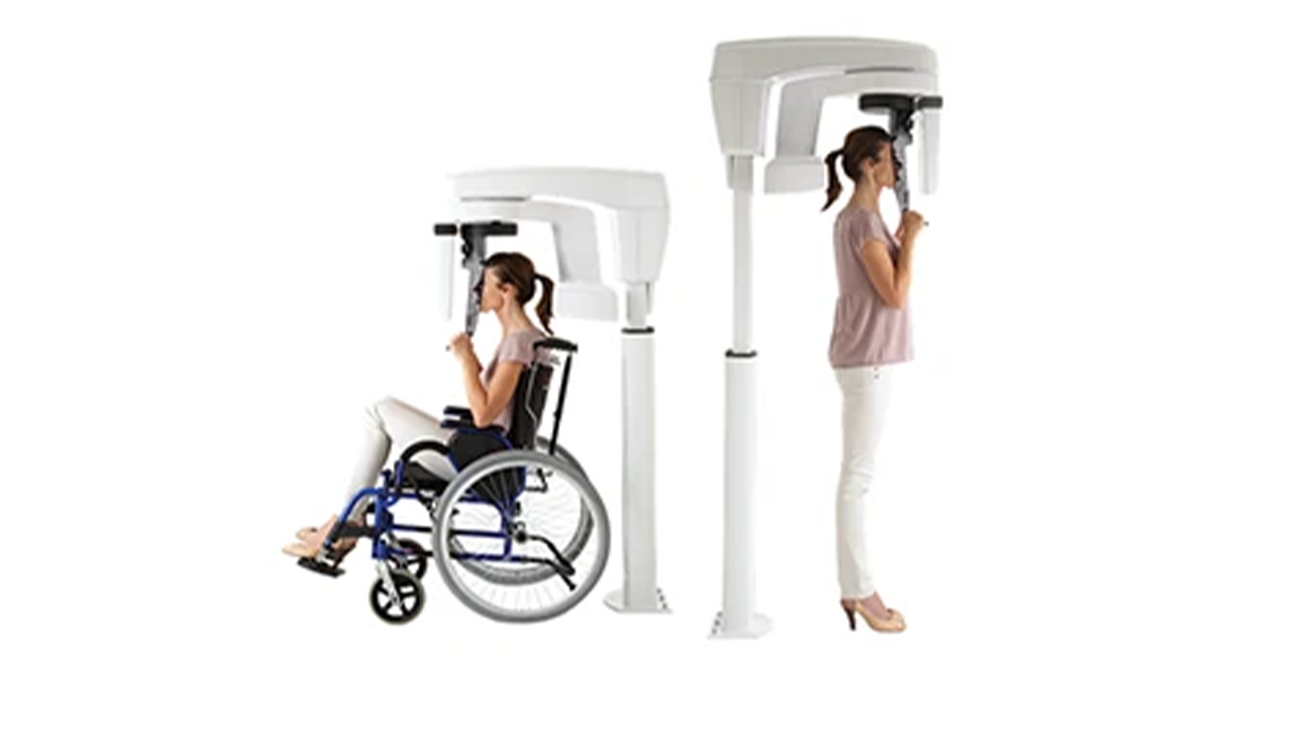
| Unit dimensions | 330 (L) x 894 (D) x 1596 (H) mm
13 (L) x 35.18 (D) x 62.83 (H) in. | ||||||||||||||||||||||||||||||||||||||||||
| Minimum required space | 1200 (L) x 1400 (D) x 2400 (H) mm
42.24 (L) x 55.11 (D) x 94.48 (H) in. | ||||||||||||||||||||||||||||||||||||||||||
| Weight | 92 kg (202 lb 13 oz) | ||||||||||||||||||||||||||||||||||||||||||
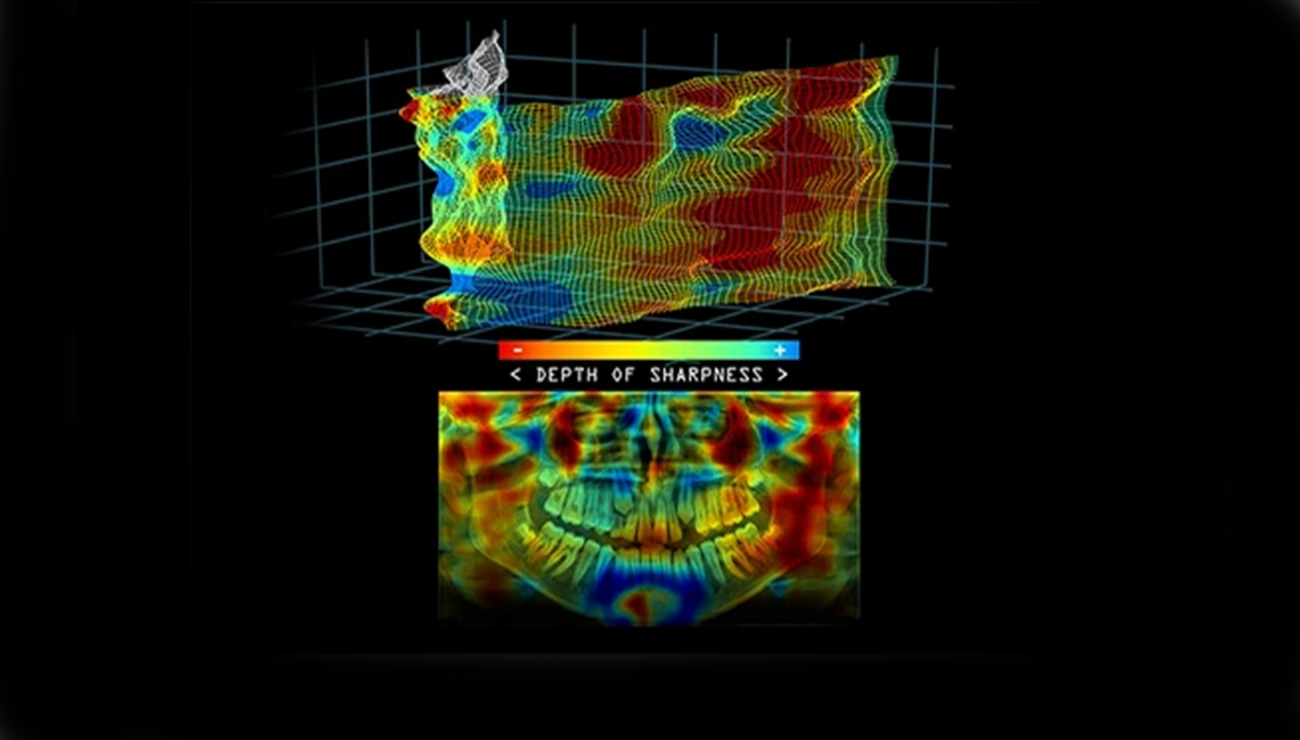
| Sensor technology | CMOS | ||||||||||||||||||||||||||||||||||||||||||
| Image field | 6.4 x 140 mm (Adult) – 6.4 x 120 mm
(Pediatric) | ||||||||||||||||||||||||||||||||||||||||||
| Gray scale | 16384 - 14 bits | ||||||||||||||||||||||||||||||||||||||||||
| Magnification | 1.2 (± 10%) | ||||||||||||||||||||||||||||||||||||||||||
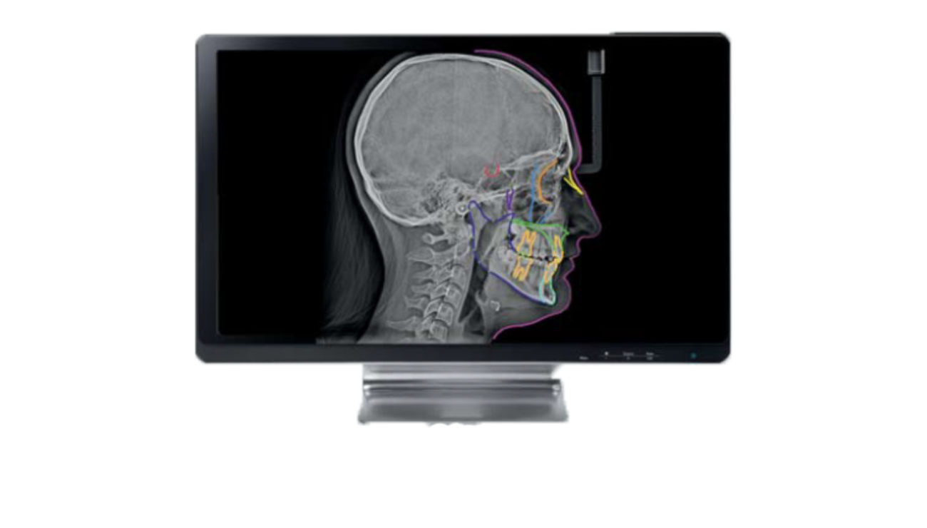
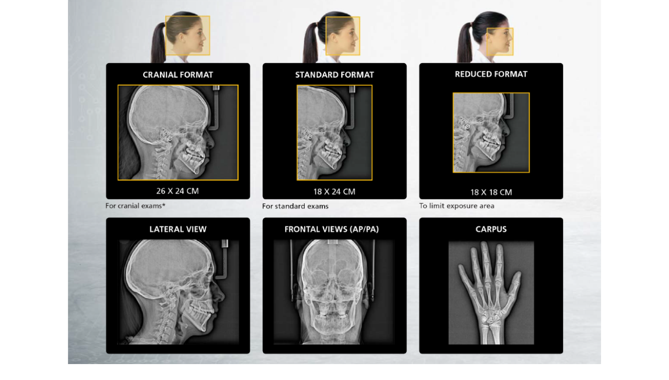
| Radiological exam options | Full panoramic, segmented panoramic,
maxillary sinus, LA TMJ x 2, LA TMJ x 4,
segmented bitewing | ||||||||||||||||||||||||||||||||||||||||||
| Exposure mode | 4 patient sizes (child, small adult,
medium adult, large adult)
3 dental arch morphologies
(normal, square, sharp) | ||||||||||||||||||||||||||||||||||||||||||
Exposure time Cephlometric Arm
| 1.98 to 14 seconds |